Precautions for mouse breeding
Ectromelia virus (ECTV) is a virus of the Orthopoxvirus genus that causes mousepox, a disease of mice. It has only been documented in mouse colonies kept for research use, but it is believed that wild populations of mice and other rodents in Europe are naturally infected with ECTV[1]. Mousepox causes skin lesions, a purulent rash on the body of mice, and generalized disease, which can be fatal. It is the only poxvirus to cause disease naturally in mice.
The reduction of viral infection in animals is one of the main points of work within the Animal Health program at Cyagen. Our mouse colonies are housed in AAALAC-accredited specific-pathogen-free (SPF) barrier facilities that guarantee exclusion of numerous pathogens, including ECTV.
The following information on animal breeding and health aims to guide investigators who are unfamiliar with mouse breeding, or who are breeding transgenic or gene-targeted mice for the first time. These suggestions are based on our experience in animal welfare and colony management; they are open to modification and should not be construed as a comprehensive set of rules.
Etiology
Classification:DNA virus, enveloped
Family:Poxviridae
Affected species: Laboratory mice, wild mice, and other wild rodents.
Frequency:Rare in laboratory mice, uncommon in wild mice.
The primary site of infection: Epidermis (Skin)
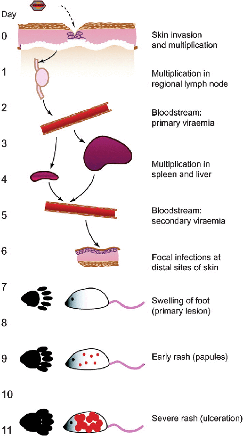
Infection process:Infection begins through a break in the skin, where the virus will replicate and then disseminate via the lymphatics. Primary viremia occurs when the virus is released into the bloodstream, which then permits infection of the spleen, liver, and other organs. Secondary viremia occurs due to the release of the virus from organs, resulting in infection of distal sites of the skin. The foot (site of primary infection) swells due to the inflammatory and immune response. In certain strains and wild mice, a rash may develop[1].

Figure 1. Sequence of events during the course of ECTV infection in a susceptible mouse following infection through the skin (footpad).[1]
Isolation:The virus may be isolated in mouse embryo cell cultures and identified by immunological means; mice are infected readily by inoculation.
Other: The natural host of ECTV (and the original source of infection in experimental mice) is not yet known.
Clinical Symptoms
Differences in resistance:
The clinical signs and development of lesions associated with mousepox depend on the background strain of mice, all of which vary greatly in sensitivity to infection. Experimental studies showed that all strains of mice were sensitive to mouse pox infection, but BALB/c, DBA/2, C3H/He mice were highly sensitive, AKR and SJL mice were moderately sensitive, and C57BL/6 mice were highly resistant to lethal infection. More resistant strains of mice may not exhibit the same clinical signs shown in other lines. Outbreaks among susceptible rats are unstable, and there are different morbidity and high mortality among susceptible mouse strains.
Features of clinical symptoms:
There can be four clinical situations which occur for an ECTV infection: acute, subacute, chronic, and asymptomatic. The acute type is seen in the first time the disease occurs in the mice population, the affected mice have a rough and dull coat, and their appetite is diminished. Acutely infected mice often die within 4-12 hours, at a mortality rate of 60-90%. Subacute infection in mice more commonly presents clinical signs in the skin, with rash, swelling, and ulceration of the skin on the nose, face, limbs and tail, accompanied by serous exudates. Often, the tail and feet become necrotic and gangrene, and the gangrene falls off and scabs in 1-2 days. The chronic type is more common in the late epidemic period, occasionally skin-type diseased mice appear and the growth of bred mice is delayed. Strains with strong resistance, such as B6, B10, and ARK, may be asymptomatic, but they will become the source of infection for other mice.

Figure 2. After depilation, mice showed a rash related to murine pox [2]
Animal Epidemiology
Propagation mode:
1. Mousepox virus can cause mousepox in mice, which can be transmitted through direct contact or vectors.
2. Exposure to viruses through skin wounds is a natural route of infection.
3. The susceptible strains are damaged 7-11 days after infection.
4. The virus is shed for 3 weeks.
5. For 16 weeks after the mouse infection, the virus can still be detected in the scabs and feces of the mouse.
6. The spread of mousepox from one cage to another is mainly caused by personnel operations, who carry the virus from infected to healthy mice.
Epidemiology of mousepox virus infection in mice with different resistance:
1. Moderately resistant mice are prone to natural transmission. Such mice survive long enough to cause skin damage and detoxify for a relatively long time. Infectious viruses persist in excreta and shed scabs, further increasing the risk of transmission. Although the virus detox period usually lasts about 3 weeks, viruses found in scabs and/or feces can survive for up to 16 weeks.
2. Resistant mouse strains are also dangerous because they can release viruses during subclinical infections. However, mice with strong resistance tend to be infected for a short period of time.
3. If eliminated and treated in time, the risk of transmission of infection by highly susceptible mice is relatively small, because these mice have died before the virus is released in large quantities.
4. Therefore, mixing infected mice with strong or moderate resistance and highly susceptible mice can cause explosive viral infections to spread.
Epidemiology of different ages or physiological periods:
1. Young and old mice are generally more susceptible than adult mice and are more likely to die after infection.
2. In the mousepox-positive breeding mouse population, maternal immunity can protect the young mice from death, but not the young mice from infection. These pups may subsequently become infected through contact exposure.
Pathology
Pathogenesis of Mousepox
Fenner's classic description of ECTV pathogenesis is still applicable (Figure 3).

Figure 3. Diagram illustrating the pathogenesis of mousepox. [3]
Following skin invasion, viral multiplication occurs in the draining lymph node and a primary viremia ensues (Fig 3). Splenic and hepatic involvement begin within 3–4 days, whereupon larger quantities of virus are disseminated in blood to the skin. This sequence takes approximately 1 week and, unless mice die of acute hepatosplenic infection, ends with the development of a primary skin lesion at the original site of viral entry. The primary lesion is due to the development of antiviral cellular immunity.
Severe hepatocellular necrosis occurs in susceptible mice during acute stages of mousepox. White spots indicative of necrosis develop throughout the liver. In nonfatal cases, regeneration begins at the margins of necrotic areas, but inflammation is variable. Splenic necrosis in acute disease commonly precedes hepatic necrosis but is equally or more severe (Fig 4). Necrosis and scarring of red and white pulp can produce a macroscopic “mosaic” pattern of white and red-brown (Fig 5). Necrosis of thymus, lymph nodes, Peyer's patches, intestinal mucosa, and genital tract also have been observed during acute infection, whereas resistant or convalescent mice can develop lymphoid hyperplasia. Severe intestinal infection may be accompanied by hemorrhage.

Figure. 4. Splenic necrosis in acute mousepox.[3]

Figure. 5. “Mosaic spleen” from a mouse that survived acute mousepox.[3]
Diagnosis
1. If the above symptoms occur in an animal facility, or if a susceptible strain has a large area of death that cannot be explained, the mousepox virus should be suspected first.
2. Serological diagnosis can be performed by MFIA/ELISA or IFA. If the animal recovers, protective antibodies will be produced.
3. The following damages were found during autopsy to indicate murine pox infection: spleen fibrosis in recovered mice; liver, spleen, and skin damage in diseased mice.
4. Observing the characteristic eosinophilic inclusion bodies in the cytoplasm helps to detect infection.
5. PCR testing of skin damage can confirm the diagnosis.
6. If immunized with a virus vaccine, it can produce false positive serological results.
Differential diagnosis
Mousepox must be distinguished from other infectious diseases with high morbidity and mortality, such as Sendai pneumonia, mouse hepatitis, and Tyzzer's disease. The skin lesions of chronic mousepox must be distinguished from other skin diseases caused by opportunistic or pathogenic bacteria, ascariasis, and bites.
Prevention and control
1. Establish an animal health monitoring system and regularly monitor animal health.
2. For foreign animals and animal-derived products (such as serum, tumors, cell lines, etc.), they must be strictly reviewed and tested when necessary in quarantine and isolation.
3. Implement protocol for foreign mice entering the facility, such as validating the health background. Prevent wild mice from entry.
4. Once the infection is confirmed, the group must be cleared, then thoroughly disinfected, and finally re-established by cesarean section or embryo transfer purification.
5. Infected animal facilities must be thoroughly cleaned and disinfected. It is best to use formalin or hydrogen peroxide gas for fumigation.
6. Mousepox virus in the blood can survive for 11 days at room temperature. All utensils in animal facilities should be immediately disposed of as a source of pollution (incineration or high pressure).
7. High pressure, formalin fumigation, and conventional disinfectants can all inactivate mousepox virus.
Experimental influence
The main threat of mousepox is the death of susceptible mice. The resulting loss of time, animal and economic costs can be huge.
Cyagen' s Animal Health program implements an array of monitoring and testing methods to maintain the optimal health of our mouse and rat colonies. All our animal colonies are housed in AAALAC-accredited specific-pathogen-free (SPF) barrier facilities that guarantee exclusion of numerous pathogens, including ECTV.
References:
[1]Esteban, David J., and R. Mark L. Buller. "Ectromelia virus: the causative agent of mousepox." Journal of General Virology86.10 (2005): 2645-2659.
[2] Laboratory Animal Medicine [M] Third Edition 2015
[3] Infectious Agent Information | Charles River (criver.com)
[4] 贺争鸣等.实验动物疫病学[M].北京.中国农业出版社2015
[5] 胡建华等,实验动物学教程[M].上海,上海科学技术出版社2009
We will respond to you in 1-2 business days.