Introduction: Acute myocardial infarction is myocardial necrosis caused by acute and persistent ischemia and hypoxia of coronary arteries, accompanied by the increase of serum myocardial enzyme activity and the abnormal characteristic electrocardiogram changes. It is normally complicated with arrhythmia, shock or heart failures, which can result in sudden death. The symptoms and signs of clinical myocardial infarction can be simulated through left coronary artery ligation.
Animal requirement: Rats and mice - males are preferable. In special cases, female animals may be used. Age and weight can be adjusted accordingly.
Brief Protocol:
- Incision of left chest, muscle separation to expose the ribs and then intercostal incisions in the fourth and fifth ribs.
- Open the chest to expose the heart. Permanently ligate the left anterior descending coronary artery with a piece of suture to generate a myocardial infarction model.
- Or ligate the left coronary artery for a certain period, then restore coronary blood flow to generate a model of ischemia-reperfusion injury.
- Close the chest incision and suture the skin incision
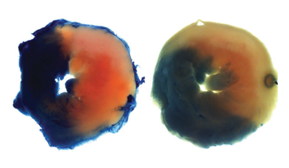 Evans Blue and TTC staining myocardial infarction mice, showed as normal myocardial perfusion area (blue), junction area (red) and the infarction area (pale white). Refer to Journal of Clinical Investigation, 2016; 126(12): 4654-4658.
 Turnaround time:
Acute and chronic experiments can be performed. Postoperative time can range from a few hours to several weeks (heart failure model).
Follow-up service:
- 1. After-surgery routine observation: daily activities and diet intake are observed by full-time veterinarians, and pain relief treatment is applied accordingly.
- 2. Cardiac ultrasonography: comprehensive evaluation of cardiac structure and function is achieved by ultra-high-definition small-animal-specific ultrasound machine. The evaluation of cardiac function before and after surgery (different periods of ventricular hypertrophy or heart failure development) includes the wall thickness (IVSs, IVSd, LVPWs, LVPWd), ventricular volume (LV Vols, LV Vold), ventricular lumen diameter (LVIDs, LVIDd), ejection fraction (EF%), fractional shortening (FS%) etc. Customized ultrasonic tests can be performed upon request.
- 3. Post-operative electrocardiogram detection: ECG records analysis at multiple time points.
- 4. Evans Blue and TTC staining myocardial infarction area analysis: For acute myocardial infarction test, double staining is adopted to distinguish normal myocardial perfusion area, infarction area and the junction area. Then, quantitative image analysis is performed.
- 5. Blood sample collection: 0.7-1.0 mL venous blood, collected from adult mice or rats according to weight, can be used for various biochemical tests. The serum or plasma is stored in a -80⁰C refrigerator until delivery or biochemical detection.
- 6. Cardiopulmonary specimen collection: Cardiopulmonary specimens are collected from different sections. The heart and lung specimens can be used to calculate the heart weight to body weight ratio and lung weight to body weight ratio. The specimens can be stored in a -80⁰C refrigerator or fixed with formalin for further pathological examination.
Animal ethical censorship and management
The Institutional Animal Care and Use Committee (IACUC) reviews the experimental protocol and manages the entire surgical procedure, including anesthesia, analgesia, animal preparation, and post-operative management.
Contact us:
For any surgical disease model, the strategy can be adjusted according to the scientific problems that researchers want to solve. If you have any special requests, please contact 86 20-31601779 or email apac-service@cyagen.com. Cyagen provides surgical model services personalized for your research goals - our senior technical team will develop a detailed experimental plan according to your needs and keep you updated throughout the model generation process.
References:
- 1. L. H. Michael, M. L. Entman, C. J. Hartley, K. A. Youker, J. Zhu, S. R. Hall, H. K. Hawkins, K. Berens, and C. M. Ballantyne. Myocardial ischemia and reperfusion: a murine model. Am J. Physiol. 1995; 269 (Heart Circ. Physiol. 38): 2147-2154.
- 2. Rongli Zhang, Douglas T. Hess, James R. Reynolds, Jonathan S. Stamler. Hemoglobin S-nitrosylation plays an essential role in cardioprotection. Journal of Clinical Investigation, 2016; 126(12): 4654-4658.

